
Introduction
The adrenal cortex is an essential component of the endocrine system, responsible for producing steroid hormones that regulate metabolism, electrolyte balance, cardiovascular function, immune responses, and sexual development. These hormones are indispensable for maintaining physiological homeostasis and enabling the body to respond appropriately to physical and psychological stress.
The adrenal glands are paired endocrine organs situated above the kidneys. Each gland consists of two anatomically and functionally distinct regions: the outer adrenal cortex and the inner adrenal medulla. While the adrenal medulla synthesizes catecholamines, primarily epinephrine and norepinephrine, the adrenal cortex produces steroid hormones derived from cholesterol.
The three principal groups of adrenal cortical hormones are:
-
Glucocorticoids, predominantly cortisol, which regulate energy metabolism, stress adaptation, immune function, and inflammatory responses.
-
Mineralocorticoids, predominantly aldosterone, which maintain sodium and potassium balance, extracellular fluid volume, and blood pressure.
-
Adrenal androgens, principally dehydroepiandrosterone (DHEA), dehydroepiandrosterone sulfate (DHEAS), and androstenedione, which contribute to androgenic activity and serve as precursors for more potent sex steroids.
Although all three hormone classes originate from cholesterol, their synthesis and secretion are controlled by distinct regulatory mechanisms. Understanding these processes provides the physiological foundation for interpreting adrenal disorders, including adrenal insufficiency, Cushing syndrome, primary aldosteronism, and congenital adrenal hyperplasia [1,2].
1. Anatomical and Functional Organization of the Adrenal Cortex
The adrenal cortex constitutes most of the adrenal gland and is organized into three histologically distinct zones. Each zone possesses a characteristic complement of steroidogenic enzymes, enabling preferential production of particular steroid hormones.
1.1 Functional zones of the adrenal cortex
|
Zone |
Principal hormones |
Main regulatory factors |
|---|---|---|
|
Zona glomerulosa (outer) |
Aldosterone |
Angiotensin II, plasma potassium; ACTH has a secondary role |
|
Zona fasciculata (middle) |
Cortisol |
Adrenocorticotropic hormone (ACTH) |
|
Zona reticularis (inner) |
DHEA, DHEAS, androstenedione |
ACTH and additional intra-adrenal regulatory factors |
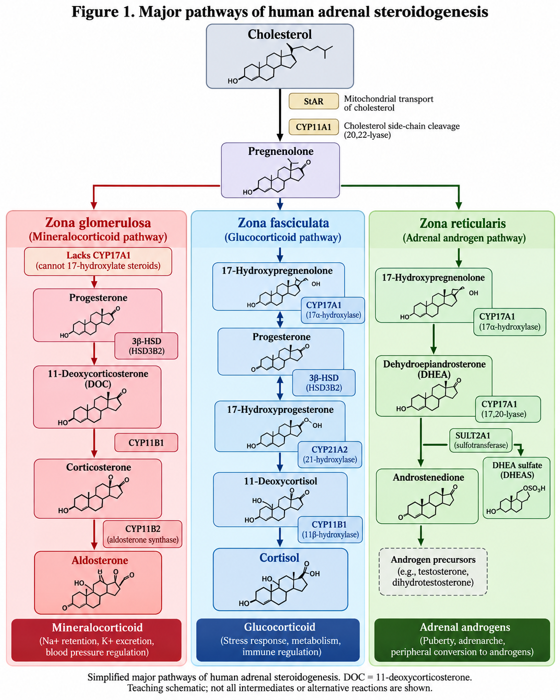
The zona glomerulosa, immediately beneath the adrenal capsule, contains cells organized into rounded clusters or arches. Its distinctive expression of aldosterone synthase (CYP11B2), together with the absence of CYP17A1, allows synthesis of aldosterone rather than cortisol.
The zona fasciculata is the largest cortical zone. It consists of lipid-rich cells arranged in cords and is the principal site of cortisol synthesis. Its cells contain the enzymes required for glucocorticoid production, including CYP17A1 and CYP11B1.
The zona reticularis lies adjacent to the adrenal medulla. Its steroidogenic machinery favors androgen precursor production, partly because of relatively high CYP17A1 17,20-lyase activity and comparatively low 3β-hydroxysteroid dehydrogenase type 2 activity.
The distinction between these zones is essential: differences in steroidogenic enzyme expression, rather than separate cholesterol precursors, determine their predominant hormonal products [1,2].
2. Biosynthesis of Adrenal Cortical Hormones
All adrenal cortical hormones are steroid compounds synthesized from cholesterol through sequential enzymatic reactions collectively termed steroidogenesis.
Unlike peptide hormones, steroid hormones are not stored in substantial quantities within secretory granules. Instead, they are synthesized largely in response to physiological stimulation and diffuse across cell membranes after production.
2.1 Cholesterol as the precursor
Cholesterol required for adrenal steroidogenesis originates primarily from circulating lipoproteins, particularly low-density lipoproteins (LDL), and from intracellular cholesterol ester stores. De novo cholesterol synthesis also contributes.
The initial steps involve:
-
Mobilization of cholesterol from intracellular stores or uptake from circulating lipoproteins.
-
Transport of cholesterol to the inner mitochondrial membrane, facilitated by steroidogenic acute regulatory protein (StAR).
-
Conversion of cholesterol into pregnenolone by the mitochondrial cholesterol side-chain cleavage enzyme, CYP11A1 (P450scc).
-
Further enzymatic conversion of pregnenolone into glucocorticoids, mineralocorticoids, or androgen precursors.
Transfer of cholesterol to the inner mitochondrial membrane is a critical acutely regulated step in steroid hormone production. The subsequent enzymatic reactions take place in both mitochondria and the smooth endoplasmic reticulum [2].
2.2 Major steroidogenic pathways
2.3 Important steroidogenic enzymes
|
Enzyme |
Principal function |
|---|---|
|
CYP11A1 |
Converts cholesterol into pregnenolone |
|
3β-HSD2 (HSD3B2) |
Converts Δ5 steroids to their corresponding Δ4 steroids |
|
CYP17A1 |
Catalyzes 17α-hydroxylase and 17,20-lyase reactions |
|
CYP21A2 |
Catalyzes 21-hydroxylation during cortisol and aldosterone precursor synthesis |
|
CYP11B1 |
Converts 11-deoxycortisol into cortisol |
|
CYP11B2 |
Catalyzes the terminal steps of aldosterone synthesis |
|
SULT2A1 |
Sulfates DHEA to form DHEAS |
The clinical importance of these enzymes becomes evident in congenital adrenal hyperplasia. For example, CYP21A2 deficiency impairs cortisol synthesis and, in severe forms, aldosterone synthesis, while diverting steroid precursors toward androgen production [2].
3. Glucocorticoids: Physiology of Cortisol
Cortisol is the principal endogenous glucocorticoid in humans. It is synthesized predominantly in the zona fasciculata and influences carbohydrate, protein, and lipid metabolism, cardiovascular responsiveness, immune function, and adaptation to stress.
Its physiological effects are both permissive and regulatory. At normal concentrations, cortisol supports essential metabolic and cellular functions, while increased secretion during stress helps maintain energy availability and circulatory stability.
3.1 Regulation of cortisol secretion: the HPA axis
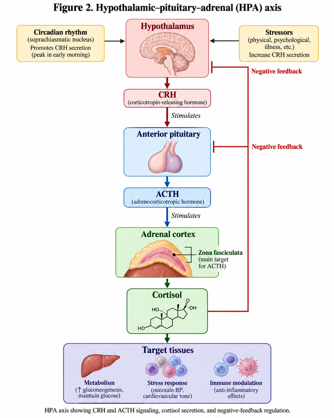
ACTH acts on melanocortin 2 receptors (MC2R) expressed on adrenal cortical cells. Activation stimulates Gs protein-dependent adenylyl cyclase activity, raising intracellular cyclic adenosine monophosphate (cAMP) and activating protein kinase A (PKA).
These signaling events rapidly increase cholesterol availability for steroidogenesis and, with sustained stimulation, modify steroidogenic enzyme expression and support adrenal cortical growth.
Cortisol subsequently suppresses hypothalamic and pituitary activity through negative feedback, thereby limiting excessive hormone secretion [1,3].
3.2 Circadian and pulsatile secretion
Cortisol secretion follows a well-defined circadian rhythm coordinated by the central biological clock and sleep–wake cycle.
In individuals following a conventional daytime schedule, cortisol levels generally rise during the latter part of sleep, peak around the early morning or shortly after awakening, and decline over the day toward a late-evening or early-night nadir.
Superimposed on this circadian pattern are ultradian pulses of ACTH and cortisol secretion. Therefore, cortisol concentrations fluctuate even within relatively short intervals.
These patterns are important when interpreting laboratory results. A cortisol concentration obtained in the early morning cannot be interpreted using the same expectations as a late-night value, and shift work may alter the usual rhythm.
The HPA axis also responds dynamically to stressors such as infection, trauma, surgery, hypoglycemia, and severe illness [1,3].
3.3 Transport and metabolism of cortisol
In circulation, most cortisol is reversibly bound to plasma proteins, particularly corticosteroid-binding globulin (CBG, also called transcortin) and albumin. A smaller free fraction is available for tissue uptake and receptor activation.
Because cortisol is lipophilic, it readily enters cells. Its local activity is further regulated by the 11β-hydroxysteroid dehydrogenase enzyme system:
-
11β-HSD1 predominantly regenerates active cortisol from cortisone in important metabolic tissues, including the liver and adipose tissue, under normal physiological conditions.
-
11β-HSD2 converts cortisol to inactive cortisone in aldosterone-sensitive epithelia, protecting mineralocorticoid receptors from excessive stimulation by cortisol.
Cortisol undergoes extensive metabolism in the liver and other tissues, with metabolites predominantly eliminated through the kidneys [1,2,5].
3.4 Physiological actions of cortisol
A. Carbohydrate metabolism
Cortisol is a major counter-regulatory hormone that helps maintain plasma glucose availability, especially during fasting and stress.
Its principal metabolic effects include stimulation of hepatic gluconeogenesis, increased availability of gluconeogenic substrates, and reduced insulin-mediated glucose uptake in certain peripheral tissues.
Cortisol also has permissive effects on other hormones involved in glucose homeostasis, particularly glucagon and catecholamines.
In physiological concentrations, these actions help prevent hypoglycemia during prolonged fasting. Persistent excess, however, can contribute to insulin resistance and hyperglycemia.
B. Protein metabolism
Cortisol promotes protein breakdown in several extrahepatic tissues, especially skeletal muscle, during catabolic conditions. The resulting amino acids provide substrates for hepatic gluconeogenesis and other metabolic processes.
Sustained glucocorticoid excess can impair protein synthesis, reduce muscle mass, weaken connective tissues, and delay wound healing.
The severity of these effects depends on circulating hormone concentrations, duration of exposure, nutritional status, and tissue-specific sensitivity.
C. Lipid metabolism
Cortisol stimulates lipolysis in certain adipose depots, particularly through permissive interactions with catecholamines and other metabolic regulators.
However, its effects on adipose tissue are complex and depend on anatomical fat distribution, insulin concentrations, and duration of exposure.
Chronic cortisol excess may promote central adiposity and ectopic lipid accumulation while contributing to adverse metabolic changes. Consequently, cortisol should not be regarded simply as a hormone that uniformly increases fat breakdown.
D. Cardiovascular regulation
Cortisol supports normal cardiovascular function by maintaining vascular responsiveness to catecholamines and other vasoactive substances.
It contributes to vascular tone, systemic blood pressure regulation, and maintenance of an adequate circulatory response during physiological stress.
Cortisol deficiency may result in reduced vascular responsiveness and hypotension. Severe deficiency, especially during acute illness, can contribute to circulatory collapse.
Conversely, prolonged cortisol excess may contribute to hypertension through multiple mechanisms, including enhanced vascular reactivity and, under some circumstances, activation of mineralocorticoid pathways.
E. Immune and inflammatory responses
Glucocorticoids influence both innate and adaptive immunity and play an important role in restricting excessive inflammatory responses.
Through glucocorticoid receptor-mediated changes in gene expression, cortisol regulates cytokine production, inflammatory cell trafficking, and the expression of numerous inflammatory mediators.
At pharmacological concentrations, glucocorticoids suppress several pro-inflammatory pathways, including aspects of NF-κB and AP-1 signaling. They also reduce leukocyte recruitment and modify the activity of lymphocytes, macrophages, and other immune cells.
Physiological cortisol concentrations have more nuanced immunoregulatory effects, and normal cortisol signaling is important for an appropriately balanced immune response.
F. Bone and connective tissue
Normal glucocorticoid signaling participates in bone development and remodeling. However, sustained glucocorticoid excess inhibits osteoblast function, reduces bone formation, and can negatively affect calcium balance and skeletal integrity.
Long-term excessive exposure increases the risk of osteoporosis and fragility fractures.
G. Central nervous system
Cortisol influences cognition, alertness, emotional processing, memory, and sleep–wake regulation through actions on glucocorticoid and mineralocorticoid receptors in the brain.
Both deficient and excessive glucocorticoid activity may disturb mood, sleep, and cognitive function. The physiological effects depend on concentration, timing, receptor distribution, and duration of exposure.
3.5 Permissive actions of cortisol
A particularly important concept in endocrine physiology is the permissive action of glucocorticoids.
A permissive action occurs when a hormone enables another hormone or signaling pathway to exert its full physiological effect without necessarily producing that effect independently.
For example, normal cortisol concentrations help preserve vascular sensitivity to catecholamines and support the metabolic responses of tissues to glucagon and other counter-regulatory signals.
This explains why glucocorticoid deficiency can cause profound physiological dysfunction even when concentrations of other regulatory hormones remain adequate.
4. Mineralocorticoids: Physiology of Aldosterone
Aldosterone is the principal mineralocorticoid hormone in humans. It is synthesized in the zona glomerulosa and plays a central role in maintaining extracellular fluid volume, blood pressure, and potassium homeostasis.
Unlike cortisol, which is principally regulated by ACTH, aldosterone secretion is controlled predominantly by the renin–angiotensin–aldosterone system (RAAS) and plasma potassium concentration.
4.1 Regulation of aldosterone secretion
A. Renin–angiotensin–aldosterone system
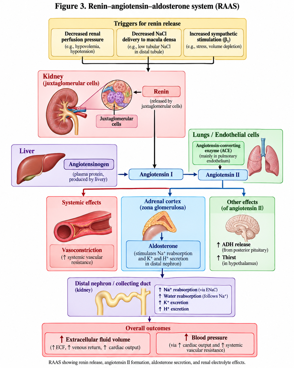
Renin is an enzyme secreted by juxtaglomerular cells in the kidneys. It cleaves circulating angiotensinogen, produced predominantly by the liver, into angiotensin I.
Angiotensin-converting enzyme subsequently converts angiotensin I into angiotensin II. This peptide stimulates aldosterone synthesis by binding to angiotensin II type 1 (AT1) receptors on zona glomerulosa cells.
Activation of these receptors increases intracellular calcium signaling, promoting cholesterol mobilization and the expression and activity of steroidogenic machinery, including aldosterone synthase.
As aldosterone increases renal sodium reabsorption, extracellular fluid volume may expand. The resulting increase in renal perfusion suppresses renin secretion, completing the regulatory feedback loop [1,6].
B. Plasma potassium concentration
Potassium is an independent and physiologically important regulator of aldosterone secretion.
An increase in extracellular potassium depolarizes zona glomerulosa cell membranes, promoting calcium entry through voltage-dependent calcium channels. The rise in intracellular calcium stimulates aldosterone synthesis.
Conversely, reduced plasma potassium generally suppresses aldosterone secretion.
This regulatory mechanism allows aldosterone to promote renal potassium elimination even when major changes in extracellular fluid volume are absent.
C. ACTH and other modulators
ACTH can acutely stimulate aldosterone secretion, but its sustained physiological control is considerably less important than that of angiotensin II and plasma potassium.
Atrial natriuretic peptide (ANP) inhibits aldosterone secretion and promotes sodium excretion during states of increased cardiac filling.
4.2 Mechanism of action of aldosterone
Aldosterone exerts its principal classical actions through the intracellular mineralocorticoid receptor (MR; NR3C2).
In the kidney, aldosterone acts particularly on principal cells of the aldosterone-sensitive distal nephron, including the late distal convoluted tubule, connecting tubule, and collecting duct.
Following receptor activation, aldosterone regulates transcription of proteins involved in sodium and potassium transport, including:
-
Epithelial sodium channels (ENaC) on the apical membrane.
-
Na⁺/K⁺-ATPase on the basolateral membrane.
-
Serum- and glucocorticoid-regulated kinase 1 (SGK1), which contributes to increased ENaC abundance and activity.
-
Components of the potassium secretory apparatus, including renal outer medullary potassium (ROMK) channels.
These coordinated effects increase sodium reabsorption and establish favorable electrical conditions for potassium secretion.
Aldosterone also influences acid–base homeostasis, partly by promoting hydrogen ion secretion through renal intercalated cell mechanisms.
4.3 Physiological actions of aldosterone
A. Sodium homeostasis
Aldosterone promotes sodium conservation by increasing sodium reabsorption in the distal nephron.
This function becomes particularly important during sodium depletion, reduced effective circulating volume, or other conditions that activate RAAS.
B. Potassium homeostasis
Aldosterone facilitates urinary potassium excretion by increasing electrogenic sodium reabsorption and supporting potassium secretion into the tubular lumen.
Its physiological importance is illustrated by aldosterone deficiency, which can result in hyperkalemia.
Conversely, excessive aldosterone activity may cause renal potassium wasting and hypokalemia, although serum potassium can remain normal in primary aldosteronism.
C. Extracellular fluid volume and blood pressure
By promoting sodium retention, aldosterone contributes to extracellular fluid volume regulation. Water balance, however, is also strongly influenced by vasopressin, thirst, and renal water handling.
Aldosterone does not simply cause unrestricted water retention. In healthy individuals, compensatory mechanisms, including pressure natriuresis, limit progressive volume expansion.
D. Acid–base balance
Aldosterone supports renal hydrogen ion secretion and acid excretion. Mineralocorticoid deficiency may contribute to hyperkalemic metabolic acidosis, whereas excessive mineralocorticoid activity may produce metabolic alkalosis.
E. Extrarenal actions
Mineralocorticoid receptors are also expressed in the cardiovascular system, brain, and other tissues.
Abnormally sustained MR activation, particularly in the setting of sodium excess, is associated with inflammatory, fibrotic, vascular, and cardiac remodeling processes [6,7].
4.4 Why does cortisol not normally dominate mineralocorticoid receptors?
An important physiological principle is that mineralocorticoid receptors bind both aldosterone and cortisol with high affinity.
Because circulating cortisol concentrations are normally much higher than aldosterone concentrations, additional mechanisms are required to ensure selective aldosterone signaling.
In classical aldosterone-sensitive epithelial tissues, 11β-hydroxysteroid dehydrogenase type 2 (11β-HSD2) converts active cortisol into cortisone, which has very low affinity for the mineralocorticoid receptor.
This allows aldosterone to act selectively at the receptor.
When 11β-HSD2 activity is deficient or inhibited, cortisol may inappropriately activate mineralocorticoid receptors, causing sodium retention, hypertension, hypokalemia, and suppression of renin and aldosterone.
This mechanism explains the syndrome of apparent mineralocorticoid excess and the potential for significant hypertension after excessive consumption of glycyrrhizin-containing liquorice products [5].
5. Adrenal Androgens: Physiology and Biological Functions
The zona reticularis produces adrenal androgen precursors, particularly DHEA, DHEAS, and androstenedione.
Compared with testosterone and dihydrotestosterone, DHEA and DHEAS possess weak direct androgenic activity. Their biological importance arises substantially from their conversion into more potent androgens and estrogens within peripheral tissues.
5.1 Synthesis and regulation
Adrenal androgen synthesis is influenced by ACTH, although its regulation is more complex than that of cortisol.
CYP17A1 possesses two enzymatic activities: 17α-hydroxylase and 17,20-lyase. The latter facilitates formation of DHEA from 17-hydroxypregnenolone.
Cytochrome b5 contributes to efficient 17,20-lyase activity, favoring androgen precursor formation in the zona reticularis.
DHEA may be converted into DHEAS through sulfation catalyzed by SULT2A1. DHEAS serves as a relatively abundant circulating reservoir of adrenal androgen precursors.
The human adrenal cortex also produces 11-oxygenated androgen precursors, which may be metabolized peripherally to potent active androgens, including 11-ketotestosterone [1,2,4].
5.2 Physiological roles of adrenal androgens
Adrenarche and pubertal development: Adrenarche is the developmental increase in adrenal androgen production that normally begins during middle childhood. It is associated with maturation of the zona reticularis and increased circulating DHEAS.
Adrenarche contributes to the development of pubic and axillary hair, adult-type body odor, and sebaceous gland activity. It is physiologically distinct from gonadarche, which involves activation of the hypothalamic–pituitary–gonadal axis.
Androgen production in females: Adrenal androgen precursors contribute to the available pool of androgens in females, including through peripheral conversion to testosterone and other bioactive steroids.
Their contribution varies with age, menopausal status, and tissue-specific steroid metabolism.
Androgen production in males: In adult males with normal testicular function, gonadal testosterone production generally dominates systemic androgen activity. Adrenal androgen precursors nevertheless remain physiologically relevant and can contribute to local tissue steroid metabolism.
Age-related changes: DHEA and DHEAS concentrations generally rise during adrenarche, reach relatively high levels in early adulthood, and progressively decline with increasing age.
Although this decline is well established, it does not itself demonstrate that DHEA replacement improves health outcomes in otherwise healthy older adults [4,8].
6. Cellular Mechanisms of Adrenal Cortical Hormone Action
The principal adrenal cortical hormones exert most of their biological effects through intracellular steroid hormone receptors belonging to the nuclear receptor superfamily.
6.1 Glucocorticoid receptors
Cortisol binds primarily to the glucocorticoid receptor (GR; NR3C1), which regulates gene expression through multiple mechanisms.
After ligand binding, receptor complexes influence transcription by interacting directly with glucocorticoid response elements and through interactions with other transcription factors.
These pathways regulate genes involved in metabolism, inflammation, cellular differentiation, and stress adaptation.
Cortisol can also act through mineralocorticoid receptors, particularly in tissues where those receptors are not protected by 11β-HSD2.
6.2 Mineralocorticoid receptors
Aldosterone binds to the mineralocorticoid receptor, regulating transcription of genes involved in electrolyte transport and other cellular functions.
Classical renal effects develop over a period of minutes to hours, with the full physiological response depending on both gene transcription and changes in transport protein activity.
6.3 Androgen receptors
Adrenal androgen precursors may undergo tissue-specific conversion to active androgens that bind the androgen receptor (AR).
Some adrenal-derived 11-oxygenated androgens possess substantial androgen receptor activity.
In addition to classical genomic signaling, steroid hormones can produce more rapid cellular responses through mechanisms that do not depend directly on new gene transcription.
7. Integrated Regulation of Adrenal Cortical Hormones
Although cortisol, aldosterone, and adrenal androgens share cholesterol as their precursor, their secretion is governed by distinct physiological requirements.
|
Feature |
Cortisol |
Aldosterone |
Adrenal androgens |
|---|---|---|---|
|
Principal origin |
Zona fasciculata |
Zona glomerulosa |
Zona reticularis |
|
Primary regulation |
HPA axis, ACTH |
RAAS and potassium |
ACTH and other factors |
|
Major physiological role |
Metabolic and stress homeostasis |
Electrolyte and volume balance |
Androgen precursor production |
|
Typical acute stimulus |
Physiological stress |
Elevated potassium or reduced effective circulating volume |
ACTH stimulation |
|
Primary receptor pathway |
GR; also MR in selected tissues |
MR |
AR/ER after conversion; some active adrenal-derived androgens act directly at AR |
These regulatory differences allow the adrenal cortex to respond appropriately to different physiological challenges.
For example, acute physiological stress generally enhances cortisol secretion to support metabolic and cardiovascular adaptation. Sodium depletion activates RAAS and stimulates aldosterone production, while hyperkalemia can independently increase aldosterone secretion.
Importantly, ACTH is not the principal sustained regulator of aldosterone production. Therefore, disorders that reduce ACTH secretion may impair cortisol production while leaving the renin–angiotensin-dependent component of aldosterone secretion relatively preserved.
8. Clinical Correlations of Adrenal Cortical Physiology
Understanding normal adrenal cortical physiology is essential for interpreting disorders involving excessive or deficient hormone secretion.
8.1 Primary adrenal insufficiency
Primary adrenal insufficiency occurs when the adrenal cortex cannot produce adequate amounts of glucocorticoids and, depending on the extent and cause of adrenal damage, mineralocorticoids.
Typical physiological consequences include impaired stress adaptation, reduced vascular responsiveness, hypotension, fatigue, and disturbances in glucose regulation.
When aldosterone secretion is significantly reduced, sodium loss, volume depletion, and hyperkalemia may also occur.
Because the defect lies in the adrenal gland, circulating ACTH is generally elevated in established primary adrenal insufficiency.
The increased ACTH and related pro-opiomelanocortin-derived peptide activity can contribute to hyperpigmentation [9].
8.2 Secondary and tertiary adrenal insufficiency
Secondary adrenal insufficiency results from insufficient pituitary ACTH secretion, while tertiary adrenal insufficiency arises from impaired hypothalamic drive or suppression of the HPA axis.
In these conditions, cortisol secretion is reduced, but aldosterone secretion is usually relatively preserved because RAAS and potassium remain its primary regulators.
This distinction explains why marked hyperkalemia and severe mineralocorticoid deficiency are more characteristic of primary adrenal insufficiency than central adrenal insufficiency.
8.3 Cushing syndrome
Cushing syndrome results from prolonged exposure to excessive glucocorticoid activity.
Its physiological manifestations reflect amplification of cortisol actions on multiple tissues and may include central adiposity, muscle weakness, impaired glucose tolerance or diabetes, hypertension, skin thinning, osteoporosis, and increased susceptibility to infection.
The syndrome may result from endogenous cortisol excess or exogenous glucocorticoid administration.
8.4 Primary aldosteronism
Primary aldosteronism is characterized by aldosterone production that is excessive relative to the body's physiological requirements and at least partly autonomous from normal regulatory mechanisms.
Its consequences include sodium retention, suppressed renin, hypertension, and increased cardiovascular risk.
Hypokalemia can occur but is not universally present; many patients have normal serum potassium concentrations.
The Endocrine Society's 2025 clinical practice guideline emphasizes recognition of primary aldosteronism as an important cause of hypertension [7].
8.5 Congenital adrenal hyperplasia
Congenital adrenal hyperplasia encompasses inherited disorders affecting enzymes or other proteins required for adrenal steroid hormone synthesis.
The most common form involves 21-hydroxylase deficiency.
When cortisol synthesis is impaired, compensatory ACTH secretion increases, promoting adrenal stimulation and accumulation of steroid precursors. Some of these precursors are diverted toward androgen synthesis.
Depending on severity, affected individuals may develop androgen excess, impaired cortisol production, and mineralocorticoid deficiency with salt wasting.
Other enzyme deficiencies produce different patterns. For example, 11β-hydroxylase deficiency can cause hypertension because of increased accumulation of mineralocorticoid-active 11-deoxycorticosterone [2].
9. Key Physiological Concepts
The most important principles can be summarized as follows:
-
The three principal adrenal cortical zones differ in their expression of steroidogenic enzymes and preferential hormone production.
-
Cholesterol is the common precursor for all adrenal cortical steroids.
-
Cortisol is principally controlled by the HPA axis and demonstrates circadian and pulsatile secretion.
-
Cortisol supports metabolic homeostasis, cardiovascular responsiveness, immune regulation, and stress adaptation.
-
Aldosterone secretion is mainly regulated by angiotensin II and potassium, rather than sustained ACTH stimulation.
-
Aldosterone promotes renal sodium conservation, potassium secretion, and acid–base homeostasis.
-
The enzyme 11β-HSD2 protects aldosterone-sensitive mineralocorticoid receptors from inappropriate cortisol stimulation.
-
Adrenal androgen precursors contribute to adrenarche and peripheral androgen and estrogen production.
-
Differences in hormone regulation explain why primary and central adrenal insufficiency produce different electrolyte and hormonal abnormalities.
Conclusion
Adrenal cortical hormones perform indispensable roles in physiological homeostasis. Cortisol regulates energy availability, vascular responsiveness, immune activity, and adaptation to stress. Aldosterone maintains extracellular fluid and electrolyte balance through coordinated renal actions, while adrenal androgens contribute to androgenic development and serve as substrates for peripheral sex steroid synthesis.
These hormones originate from a common cholesterol precursor but are synthesized in specialized cortical zones whose enzyme expression determines the final steroid products. Their secretion is regulated through complementary endocrine systems, particularly the HPA axis, RAAS, and plasma potassium-dependent signaling.
A sound understanding of these mechanisms is necessary for interpreting the pathophysiology of adrenal insufficiency, Cushing syndrome, primary aldosteronism, and congenital adrenal hyperplasia. It also provides the foundation for understanding the pharmacological actions of glucocorticoids, mineralocorticoid receptor antagonists, and inhibitors of steroid hormone synthesis.
References
Vancouver style. The references below are authentic scientific publications, clinical practice guidelines, or established academic textbook chapters. DOI and NCBI links are supplied where applicable.
1. Nicolaides NC, Willenberg HS, Bornstein SR, Chrousos GP. Adrenal cortex: embryonic development, anatomy, histology and physiology. In: Feingold KR, Adler RA, Ahmed SF, et al., editors. Endotext [Internet]. South Dartmouth (MA): MDText.com, Inc.; 2000– [updated 2023 Jun 12; cited 2026 Oct 9]. Available from: https://www.ncbi.nlm.nih.gov/books/NBK278945/
2. Miller WL, Auchus RJ. The molecular biology, biochemistry, and physiology of human steroidogenesis and its disorders. Endocr Rev. 2011;32(1):81–151. doi: 10.1210/er.2010-0013.
3. Angelousi A, Margioris AN, Tsatsanis C. ACTH action on the adrenals. In: Feingold KR, Adler RA, Ahmed SF, et al., editors. Endotext [Internet]. South Dartmouth (MA): MDText.com, Inc.; 2000– [updated 2020 Jun 13; cited 2026 Oct 9]. Available from: https://www.ncbi.nlm.nih.gov/books/NBK279118/
4. Antoniou-Tsigkos A, Zapanti E, Ghizzoni L, Mastorakos G. Adrenal androgens. In: Feingold KR, Adler RA, Ahmed SF, et al., editors. Endotext [Internet]. South Dartmouth (MA): MDText.com, Inc.; 2000– [updated 2019 Jan 5; cited 2026 Oct 9]. Available from: https://www.ncbi.nlm.nih.gov/books/NBK278929/
5. Stewart PM. Cortisol as a mineralocorticoid in human disease. J Steroid Biochem Mol Biol. 1999;69(1–6):403–408. doi: 10.1016/S0960-0760(99)00072-2.
6. Papadopoulou-Marketou N, Vaidya A, Kaltsas G, Chrousos GP. Hyperaldosteronism. In: Feingold KR, Adler RA, Ahmed SF, et al., editors. Endotext [Internet]. South Dartmouth (MA): MDText.com, Inc.; 2000– [updated 2026 Feb 16; cited 2026 Oct 9]. Available from: https://www.ncbi.nlm.nih.gov/books/NBK279065/
7. Adler GK, Stowasser M, Correa RR, Khan N, Kline G, McGowan MJ, et al. Primary aldosteronism: an Endocrine Society clinical practice guideline. J Clin Endocrinol Metab. 2025;110(9):2453–2495. doi: 10.1210/clinem/dgaf284. Published correction: J Clin Endocrinol Metab. 2025;110(11):e3933–e3934.
8. Papadopoulou-Marketou N, Kassi E, Chrousos GP. Adrenal androgens and aging. In: Feingold KR, Adler RA, Ahmed SF, et al., editors. Endotext [Internet]. South Dartmouth (MA): MDText.com, Inc.; 2000– [updated 2023 Jan 18; cited 2026 Oct 9]. Available from: https://www.ncbi.nlm.nih.gov/books/NBK279006/
9. Bornstein SR, Allolio B, Arlt W, Barthel A, Don-Wauchope A, Hammer GD, et al. Diagnosis and treatment of primary adrenal insufficiency: an Endocrine Society clinical practice guideline. J Clin Endocrinol Metab. 2016;101(2):364–389. doi: 10.1210/jc.2015-1710.
Frequently Asked Questions
What are the three main adrenal cortical hormones?
The three principal groups are glucocorticoids (mainly cortisol), mineralocorticoids (mainly aldosterone), and adrenal androgens (including DHEA, DHEAS, and androstenedione).
Which adrenal cortical hormone regulates blood pressure?
Both cortisol and aldosterone contribute to blood pressure regulation, but through different mechanisms. Aldosterone primarily influences sodium balance and extracellular fluid volume, while cortisol supports vascular responsiveness to catecholamines and other vasoactive substances.
Why is cortisol known as the stress hormone?
Cortisol secretion increases in response to many physiological stressors. It supports glucose availability, vascular stability, and coordinated metabolic and immune responses, enabling adaptation to increased physiological demands.
What is the difference between ACTH and aldosterone regulation?
ACTH is the dominant pituitary regulator of cortisol production. Aldosterone is regulated principally by the renin–angiotensin system and plasma potassium, although ACTH can exert transient stimulatory effects.
Why does adrenal insufficiency cause hypotension?
Cortisol deficiency reduces normal vascular responsiveness to vasoactive signals. When mineralocorticoid deficiency also occurs, renal sodium loss and reduced circulating volume further contribute to hypotension.